
FIG 1.
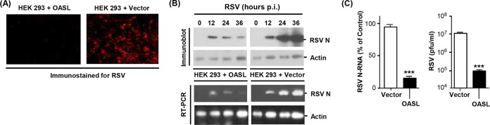
Inhibition of RSV replication in cells expressing human OASL. (A) HEK293 cells, stably transfected with V5-tagged OASL expression plasmid or the empty vector (pcDNA), as described before (18), were grown in monolayers on coverslips and infected with RSV Long at a multiplicity of infection of 3. At 18 h postinfection, cells were fixed and immunostained with mouse anti-RSV nucleoprotein (N) antibody (Abnova clone B023), followed by Alex Fluor 610-conjugated donkey anti-mouse IgG (Life Technologies). Images were captured in a Nikon AIRSI spectral confocal microscope system. (B) (Top) The same cell lines were infected as described above, and the total cell lysates were analyzed by immunoblotting using the same primary antibody described above and horseradish peroxidase (HRP)-conjugated secondary antibody, followed by ECL (enhanced chemiluminescence) detection. Actin is the loading control. (Bottom) Total RNA isolated from parallel cultures was subjected to quantitative reverse transcription-PCR (qRT-PCR), as described previously (38). The primers, synthesized by Integrated DNA Technologies (Coralville, IA), were as follows. RSV N gene, forward 5′-TGCAGGGCAAGTGATGTTAC-3′, and reverse, 5′-TTCCATTTCTGCTTGCACAC-3′; actin, forward, 5′-AGAAAATCTGGCACCACACC-3′, and reverse, 5′-GGGGTGTTGAAGGTCTCAAA-3′. A portion of the PCR sample was analyzed on 1.5% agarose gel, and the ethidium bromide-stained bands are shown. (C) (Left) Plot of the PCR results described in panel B. An average from three data sets with error bars is shown. (Right) The liberated virus in the medium at 24 h postinfection was assayed for PFU on HEp-2 monolayers as described previously (39). The asterisks indicate significance (P < 0.001).