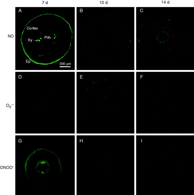
Fig. 8.
Representative images of transverse sections of hypocotyl of pepper seedlings illustrating confocal laser scanning microscopy (CLSM) detection and visualization of endogenous nitric oxide (NO), superoxide radical (O2·–) and peroxynitrite (ONOO–) at different stages of development (7, 10 and 14 d old). (A–C) Detection of endogenous NO using diaminofluorescein-FM diacetate as a fluorescent probe. (D–F) Detection of endogenous O2·– using dihydroethidium as a fluorescent probe. (G–I) Detection of ONOO– using 3′-(p-aminophenyl) fluorescein as a fluorescent probe in transverse sections. Strong and bright green fluorescence correspond to each specific fluorescent probe in the corresponding panels. Each picture was prepared from 30 sections, which were analysed by CLSM. The orange–yellow colour corresponds to autofluorescence. En, endodermis; Ep, epidermis; Xy, xylem.

