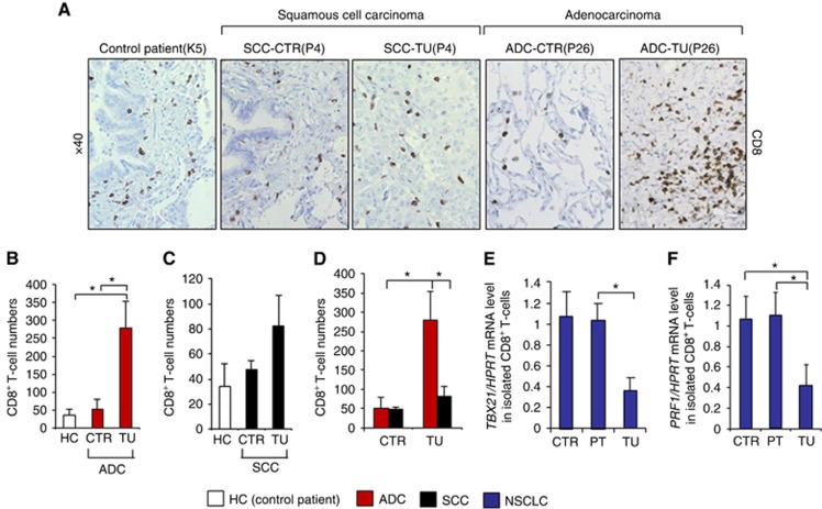
Figure 3.
Increased numbers of CD8+ T cells in the tumoral lung region of patients with adenocarcinoma. (A) IHC staining of CD8+ cells was performed on paraffin-embedded tissue sections from the control and the tumoral region of the lungs of patients with squamous cell carcinoma (n=4) and adenocarcinoma (n=4) as well as from the lungs of control subjects without lung tumour (n=3). (B–D) Bar charts represent CD8+ cell numbers counted on the tissue sections from the respective NSCLC patients as well as healthy controls. (E) qPCR-based expression analysis of TBX21 mRNA in CD8+ T cells, isolated from lung tissue samples of the tumoral, peri-tumoral and control region of patients with NSCLC (NControl=4, NPeri-tumoral=4, NTumoral=4). (F) qPCR-based expression analysis of PRF1 mRNA in CD8+ T cells, isolated from lung tissue samples of the tumoral, peri-tumoral and control region of patients with NSCLC (NControl=3, NPeri-tumoral=3, NTumoral=3). Data are shown as mean values±s.e.m. using Student's t-test *P=0.05.

