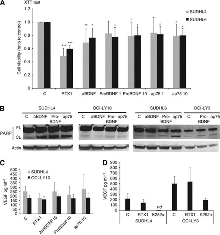
Figure 4.
A BDNF neurotrophin axis is functional in DLBCL cell lines. Effect of neutralisation of endogenous BDNF (with anti-BDNF, 15 μg ml−1) or p75NTR signalling (with anti-p75NTR neutralising antibodies, 1 and 10 ng ml−1) and exogenous pro-BDNF (1 and 10 ng ml−1) was analysed on DLBCL cells during 72 h. (A) Viability of cells exposed to anti-BDNF or pro-BDNF or anti-p75NTR was evaluated in reference to rituximab response (1 μg ml−1) using the XTT metabolic test. Data obtained on GCB DLBCL cells after 72 h exposure are done as means±s.d. of relative cell viability (ratio) related to control obtained from seven independent experiments. Student t-test was used to determine significance, *P<0.05; **P<0.01 and ***P<0.001 when compared to controls (C). (B) Analysis of apoptosis revealed by western blotting detection of PARP cleavage. Data obtained for GCB cell lines (SUDHL4 and SUDHL6) compared to ABC cell lines (OCI-LY10 and OCI-LY3) are representative of three independent experiments. Actin is done as loading control. (C) VEGF secretion by DLBCL cells was quantified by ELISA in cell supernatants after 48 h. Results are expressed as means±s.d. (pg ml−1) of at least four independent assays. **P<0.01 when compared with controls (Student t-test). (D) Effect of Trk inhibition by K252a exposure was also analysed on VEGF secretion as compared to rituximab (1 μg ml−1) in SUDHL4 and OCI-LY3 supernatants. Results are expressed as means±s.d. (pg ml−1) of three independent assays. nd= non detectable.

