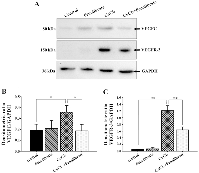
Figure 3.
Western blot analysis of vascular endothelial growth factor C (VEGFC) and VEGF receptor-3 (VEGFR-3) expression in retinal pigment epithelial cells (RPE cells). (A) Western blots representing each group: VEGFC (80 kDa), VEGFR-3 (150 kDa) and glyceraldehyde 3-phosphate dehydrogenase (GAPDH) (36 kDa). (B) Relative expression of GAPDH and VEGFC in RPE cells. (C) Relative expression of GAPDH and VEGFR-3 in RPE cells. Results shown are the means ± SD; n=8. VEGFC was expressed in normal RPE cells, whereas VEGFR-3 was not. Compared with the control group, the effects of fenofibrate on VEGFC and VEGFR-3 expression in normal RPE cells were not significant. The intracellular VEGFC and VEGFR-3 expression in RPE cells after CoCl2-induced hypoxia was significantly increased, and fenofibrate significantly reduced the synthesis of VEGFC and VEGFR-3 in RPE cells following exposure to hypoxia. *p<0.05 and **p<0.01.

