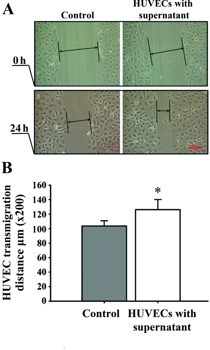
Figure 6.
Analysis of the migration of human umbilical vein endothelial cells (HUVECs) by scratch-wound assay. (A) Migration of HUVECs cultured for 24 h in the supernatant of control retinal pigment epithelial cells (RPE cells) (left panel) and in the supernatant of RPE cells epxosed to hypoxia (right panel) (x200 magnification). (B) HUVEC migration rate. The scratch width of each group was 200 µm, and the cell migration distance of the cells between the scratch edges was observed after 24 h. Results shown are the means ± SD; n=8. Compared with the control group, cutlure with the supernatant from hypoxic RPE cells significantly increased the HUVEC migration rate. *p<0.05.

