Figure 7.
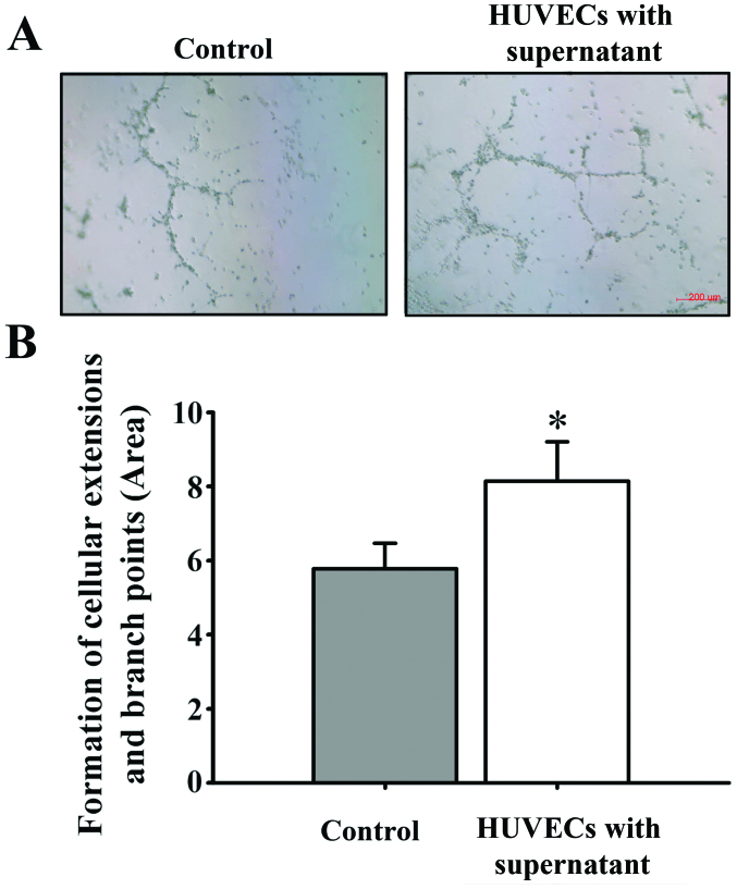
Effects of culture with the supernatant of retinal pigment epithelial cells (RPE cells) on angiogenic activity of human umbilical vein endothelial cells (HUVECs). Cultured HUVEC cells were divided into 2 groups and plated in Matrigel. The control group was treated with Dulbecco's modified Eagle's medium (DMEM) containing 1% fetal bovine serum (FBS). The experimental group was treated with the supernatant of RPE cells exposed to hypoxia. (A) Control group (left panel) and the experimental group (right panel) after 48 h of incubation (x50 magnification). (B) Area covered by cellular extensions linking HUVEC masses and branch points. These calculations were performed on images of standardized fields from at least 5 wells/experimental condition. Results are the means ± SD, with 8 samples in each group. Compared with the control group, culture with the supernatant of RPE cells exposed to hypoxia significantly increased the tube formation ability of the HUVECs. *p<0.05.

