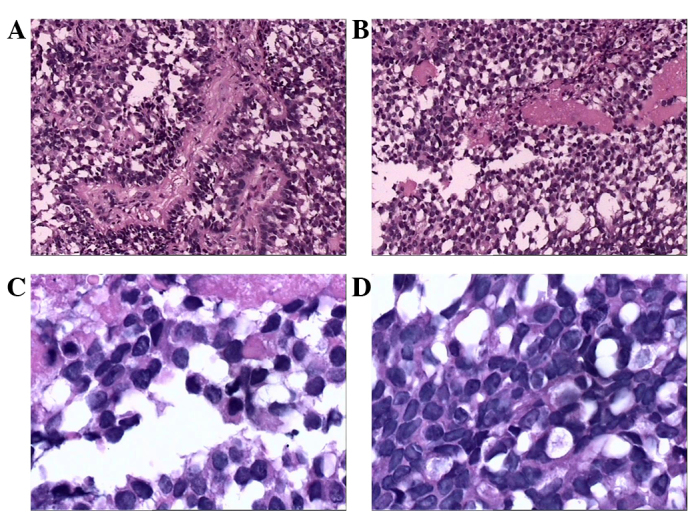
Figure 2.

Hematoxylin and eosin staining revealing the papillary features of the tumor cells. (A and B) Sections of the tumor exhibited papillary structures and a palisade arrangement surrounding the vascular pseudo-stratified columnar epithelium (magnification, x100). (C and D) The cytoplasm was hyperchromatic and exhibited irregular nuclei (magnification, x400).
