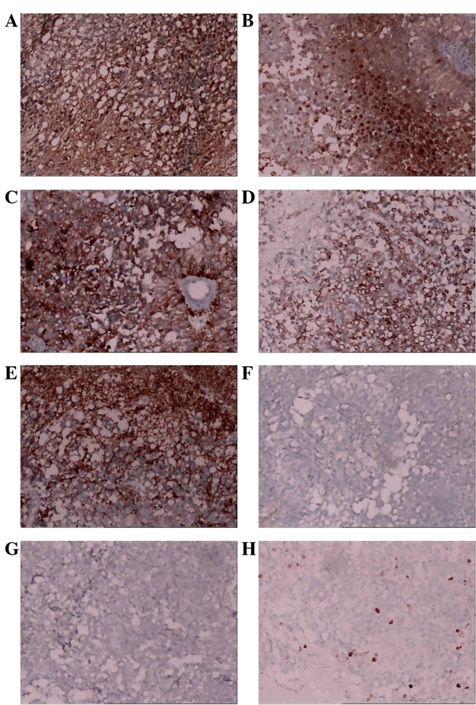
Figure 3.

Micrographs demonstrating positive immunohistochemical staining for (A) S-100, (B) neuron-specific enolase, (C) Cam5.2, (D) CK8-18 and (E) Syn, and negative staining for (F) glial fibrillary acidic protein and (G) epithelial membrane antigen. (H) Approximately 5% of the tumor cells were positive for Ki67.
