
Figure 4.
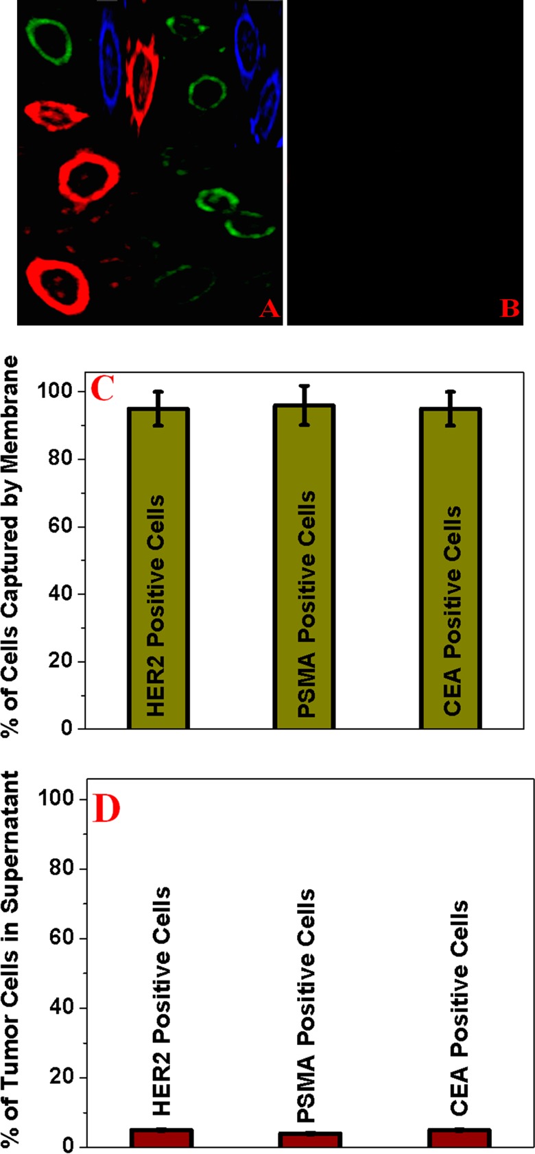
(A) Fluorescence image showing that the bioconjugated porous graphene oxide membrane is capable of capturing different types of tumor cells from infected blood. (B) Fluorescence image demonstrating that no cells are captured when a normal skin HaCaT cell is used. (C) Number of HER2 positive, PSMA positive, and CEA positive cells in membranes. (D) Number of HER2 positive, PSMA positive, and CEA positive cells in the supernatant.