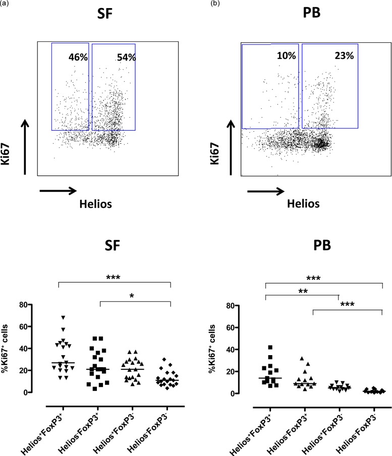
Figure 5.

Synovial Helios+FoxP3+ T cells displayed the highest frequency of cycling cells. Mononuclear cells from inflamed joints [synovial fluid (SF)] (n = 19) [13 ankylosing spondylitis (SpA), three rheumatoid arthritis (RA), three juvenile arthritis (JIA)] and from peripheral blood (PB) (n = 13) (10 SpA, three RA) of arthritis patients were analysed by flow cytometry for the proliferation marker Ki67. CD14– single cells were gated via CD3 and CD4 and subsequently Helios+FoxP3+ and Helios−FoxP3+ T cells positive for Ki67 are depicted. The dot-plots show representative Ki67 and Helios stainings of CD3+CD4+FoxP3+ cells in SF (a) and PB (b) from patients. Summary graphs of the proliferation in the different Helios FoxP3 subpopulations are shown for SF (a) and PB (b). The proliferation data were compared using the Kruskal–Wallis test together with Dunn’s multiple-comparision post-test, significance: ***P < 0·001, **P < 0·005 and *P < 0·05.
