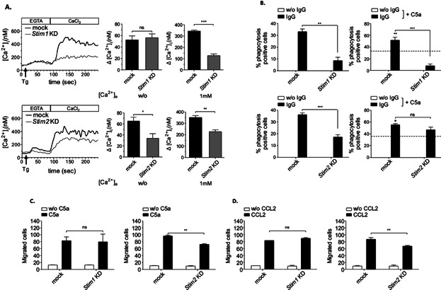
Figure 8.

Confirmative evidence for alternate functions of STIM1 and STIM2 that are differentially defective in Stim1 and Stim2 KD macrophages. (A) Stim1 and Stim2 KD RAW264.7 cells and mock-transfected controls were loaded with Fura2/AM, stimulated with 2 μM TG in EGTA-containing buffer (w/o [Ca2+]e) followed by the addition of CaCl2 (1 mM [Ca2+]e) and monitoring of [Ca2+]i. Representative measurements (upper/lower left panels) and maximal (Δ[Ca2+]i ± SEM) values (n = 5 per group) in the absence (w/o [Ca2+]e) (upper/lower middle panels) and presence (1 mM [Ca2+]e) of extracellular Ca2+ (upper/lower right panels) are shown (*P < 0.05; **P < 0.01; ***P < 0.001). Only Stim2 KD cells show a significant defect of TG-induced Ca2+ ER store release, whereas SOCE appears much lower in Stim1 KD cells. (B) The indicated cells were incubated with uncoated (w/o IgG) or IgG-coated MRBCs and percentage of phagocytosis was assessed (upper/lower left panels). Phagocytosis of IgG-opsonized MRBCs by mock-transfected cells (indicated by the intermittent line) was increased in the presence of 50 ng/mL of C5a (IgG + C5a; significance is shown in bold) in Stim2 but not Stim1 KD RAW264.7 cells (upper/lower right panels). (C, D) Cells were assayed for C5a- and CCL2-induced chemotaxis in Transwell migration assays, and migrated cells were counted by light microscopy. (B–D) The results are expressed as mean ± SEM of four to five independent experiments (**P < 0.01; ***P < 0.001).
