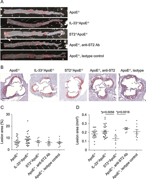
Figure 1.

Development of atherosclerosis in mice deficient in IL‐33/ST2 signaling. Representative photographs (A, B) and quantitative analysis (C, D) of atherosclerotic lesion areas in thoracic‐abdominal aortas (A, C) and aortic sinuses (B, D) of ApoE−/− (n = 20), IL‐33−/−ApoE−/− (n = 25‐26), ST2−/−ApoE−/− (n = 9), or ApoE‐/‐ mice injected with either a blocking anti‐ST2 (n = 9) or the isotype control antibody (n = 10) after 10 weeks of a cholesterol‐rich diet. All samples were stained with Oil Red O (red) for lipids and sections of aortic sinuses were counterstained with hematoxylin (purple). The extent of atherosclerotic lesions was analyzed by computer‐assisted image quantification as percentage of lipid deposition within the total surface area of the thoracic‐abdominal aortas (C) or as absolute values of lipid deposition in aortic sinuses (D), respectively. Data are shown as the mean ± SEM, 1‐way ANOVA, and Tukey's post‐test for pairwise comparison. Scale bar: 100 μm.
