. 2015 May;48(5):283–288. doi: 10.5483/BMBRep.2015.48.5.190

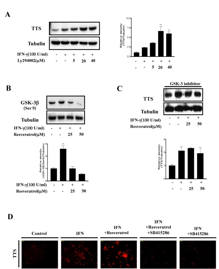
Fig. 2. GSK-3β plays a key role regulating TTS. (A), BMDCs were pretreated for 30 min in the absence or presence of a specific PI3K inhibitor (LY294002; 25-50 μm) and incubated with IFN-γ (100 U/ml) for 18 h. The mean and standard error values shown represent two independent experiments. * and ** represent significant differences at P < 0.05 and P < 0.01, respectively, compared with unstimulated cultures. (B), BMDCs were preincubated for 1 h in the absence or presence of resveratrol (25-50 μm) and stimulated with IFN-γ (100 U/mL) for 30 min. ** significant difference at P < 0.01, compared with unstimulated cells. The results are representative of two independent experiments. (C), BMDCs were pretreated for 30 min in the presence of a GSK-3 specific inhibitor (SB415286), treated with resveratrol (25-50 μm) for 1 h, and incubated with IFN-γ (100 U/ml) for 18 h. The protein levels in the cell lysates were analyzed by Western blot using the indicated antibodies. The mean and standard error values shown represent two independent experiments. (D), BMDCs were pretreated with or without a GSK-3 inhibitor (SB415286) for 30 min, treated with resveratrol (25-50 μm) for 1 h, and then incubated with IFN-γ (100 U/ml) for 18 h. The cells were fixed in 4% paraformaldehyde for 10 min, stained with mouse anti-TTS antibodies overnight at 4℃, and then stained with Alexa568-conjugated anti-mouse antibodies for 1 h at room temperature. Cell morphology and fluorescence intensity were analyzed using a Zeiss LSM510 Meta confocal laser scanning microscope. Results are representative of three independent experiments.

Fig. 2.