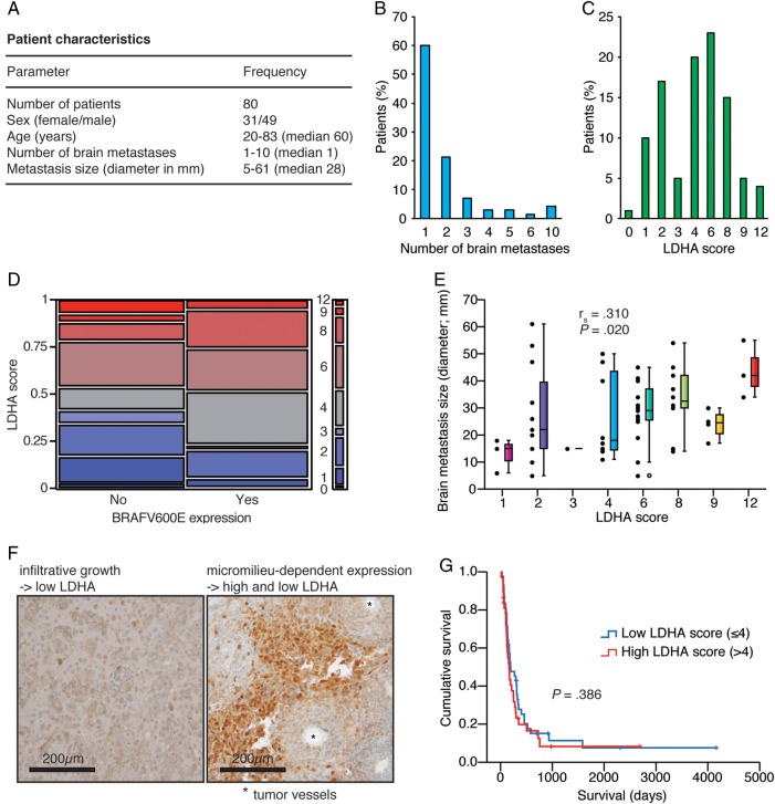
Fig. 2.
LDHA expression levels of human melanoma brain metastases was not predictive of tumor burden or survival but increased with tumor size. (A) Cohort characteristics. (B) Number of brain metastases. (C) LDHA scores. (D) BRAFV600E expression status vs LDHA score (Pearson chi-square test, P = .221). (E) LDHA score vs brain metastasis size (n = 56; Spearman correlation). (F) LDHA immunohistochemistry of diffusely infiltrating tumor cells (left) and micromilieu-dependent expression in the tumor core (right). (G) Kaplan–Meier survival plot of low vs high LDHA score (median split; Mantel–Cox log-rank test). Five patients in the low group and 7 patients in the high group were still alive or lost to follow-up.

