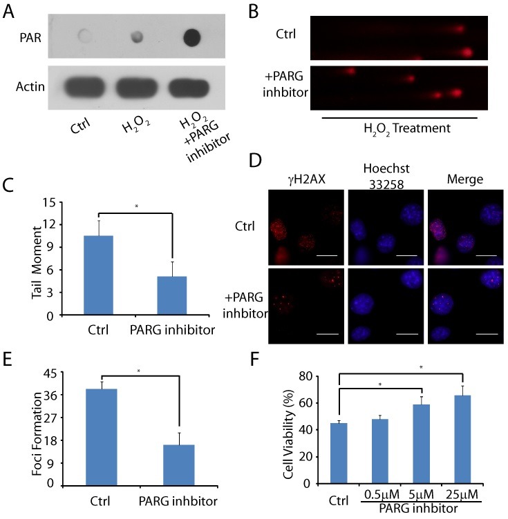
Fig. 4. PARG inhibitor reduced oxidative DNA damage level and cell death. (A) Following PARG treatment, PAR synthesis in MOVAS was examined by dot blot. (B, C) MOVAS were pre-treated with or without PARG inhibitor followed by H2O2. DNA breaks were examined at 20 min following H2O2 treatment by alkaline comet assays. Tail moment was measured. (D, E) MOVAS were pre-treated with or without PARG inhibitor. H2O2-induced DSBs were examined and the foci of γH2AX in each cell were counted (n = 15). (F) MOVAS were pre-treated with or without PARG inhibitor and then exposed to 100 ㎛ H2O2. Cell viability was evaluated using an MTT assay. The error bars represent the standard deviation, *P < 0.05.

