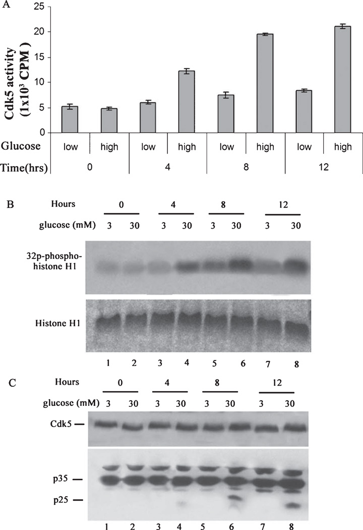
Fig. 1.
High glucose induces p25 expression and overactivates Cdk5 activity in cortical neurons, a time course study. Primary cortical neurons were treated with low (3 mM) and high (30 mM) glucose for various times. Cells were lysed and Cdk5 was immunoprecipitated using C-8 antibody from equal amounts of lysates. Immunoprecipitates were then subjected to kinase assay with histone H1 as substrate. The activity was quantified from three separate experiments as summarized in the bar graphs (A). Samples of the reactions were electrophoresed and prepared as autoradiographs as shown in (B). In separate experiments, cortical neurons were treated as above followed by SDS-PAGE and western analysis with the Cdk5 and p35 antibodies (C).

