Fig. 2.

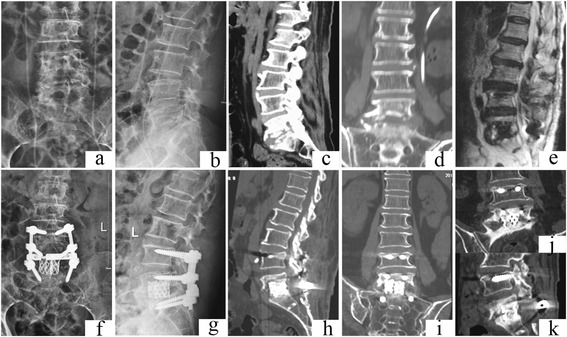
Case number 11. Preoperative image evaluation. a, b Radiograph showing L5–S1 involvement. c–d Computed tomography (CT) and e magnetic resonance imagery (MRI). f–j Postoperative radiography and h–i CT. j, k At final follow-up, the internal fixation was in good shape and interbody fusion had been obtained, without signs of tuberculosis recurrence
