
Fig. 4.
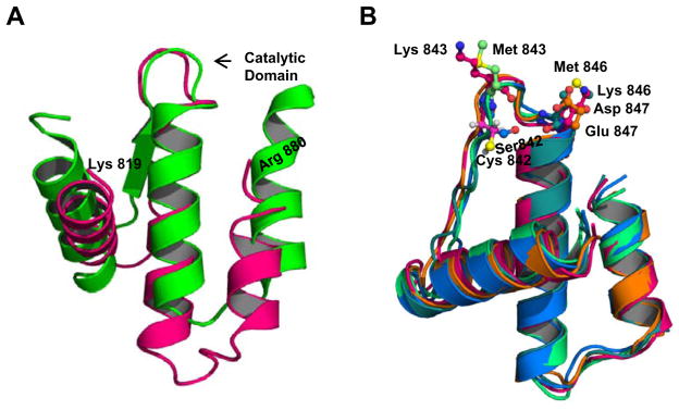
Predicted structure of the INPP4B phosphatase domain does not change with introducing mutations into catalytic loop. (A) Alignment of crystal structure models of INPP4B (pink) and PtpB (green), a protein tyrosine phosphatase from Mycobacterium tuberculosis. (B) Alignment of WT INPP4B and mutant INPP4B proteins; mutations do not alter the general orientation of adjacent helices.