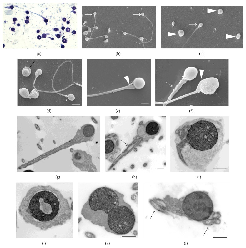
Figure 1.
Optical and ultrastructural images of round-headed sperm. (a) Thin section, optical microscopy; ((b), (c), and (d)) SEM view showing round-headed sperm (arrows) and some apparently without tail (arrowheads); (e) a conical neck (arrowhead); (f) a thick midpiece (arrow) and a cord structure at the head side (arrowhead); ((g), (h)) round heads and nuclei, anomalous axoneme in the midpiece (arrow); (i) a bending tail; (j) a detached nuclear envelope and a lacunar chromatin defect; (k) a binucleated head; (l) five axoneme sections enclosed in the same head. ((b) and (c)) White scale bars = 5 μm; (d) white scale bar = 3 μm; ((e) and (f)) white scale bars = 1 μm; ((g), (h), (j), (k), and (l)) black scale bars = 1 μm.

