Fig. 3.

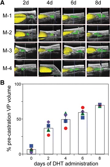
Ventral prostate lobe re-growth in previously castrated mice, following administration of exogenous DHT. a Amira generated 3D volume reconstructions from ultrasound images, of four mice (M-1 through M-4), acquired 2, 4, 6 and 8 days following the administration of DHT to the cohort of day 14 castrated mice shown in Fig. 2 (corresponding to the mice in the right-most column of B). Segmentation of the bladder (yellow) and the ventral prostate (green) is illustrated. b Plot of ventral prostate volume, following DHT administration, over time (days). Volumes determined at each time point were normalized to the pre-castration (intact) volume (in Fig. 2). Columns denote the mean and error bars correspond to the SEM. Symbols correspond to M-1 (circle); M-2 (diamond); M-3 (square) and M-4 (triangle).
