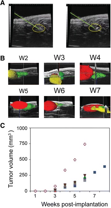
Fig. 6.

Monitoring growth of orthotopically implanted human prostate cancer xenograft. a Ultrasound image demonstrating 30-gauge needle injection (needle track is above and to the right green line) of 106 CWR22Rv1 castration resistant prostate cancer cells into the murine anterior prostate lobe (yellow outline), before (left) and after (right) injection of the cells. b Amira generated 3D volume reconstructions from ultrasound images over time (weeks). Segmentation of the xenograft tumor in the anterior prostate (red), the bladder (yellow) and the ventral prostate (green) is illustrated. Images in upper three panels were acquired with a 704 probe (80 mm FOV); lower images were acquired with a 710 probed (120 mm FOV). c Plot of orthotopic tumor volume increase over time (age, in weeks). Symbols correspond to the same animal imaged serially.
