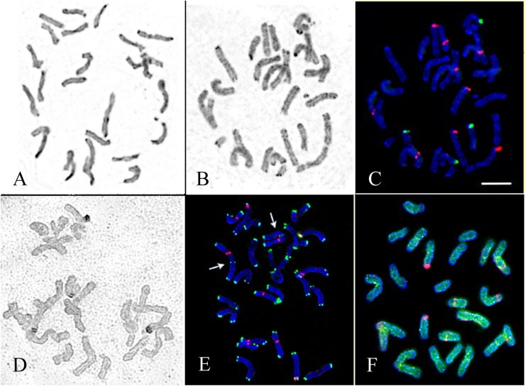
Fig 1. Chromosome spreads of D. antarctica.
(A) Giemsa C-banded chromosomes of the specimen from Galindez Island. (B) Inverted image of the DAPI/C-banded karyotype and (C) localization of 45S (green) and 5S (red) rDNA sites on chromosomes of the specimen from Galindez Island. (D) Ag-NOR staining patterns (dark segments) of chromosomes of the specimen from Skua Island. (E) Localization of telomeric repeats (green), 45S (green) and 5S (red) rDNA loci and in the karyotype of the specimen from Skua Island. Arrows point to the intercalary loci of telomere repeats detected on the largest chromosome pair. (F) Distribution of 5S rDNA sites (red) and GAA microsatellite sequence (green) on chromosomes of the specimen from Skua Island. Scale bar—5 μm.

