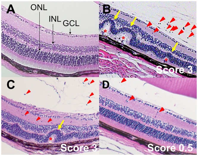
Fig 3. Representative histological findings for EAU mice.

Sections of the eye from naive mice (A) as well as from EAU mice at 21 days after injection with hIRBP(1–20) and maintenance on a control (B), ω-6 LCPUFA (C), or ω-3 LCPUFA (D) diet were stained with hematoxylin-eosin. The histological score for EAU determined from the histological findings is indicated. GCL, ganglion cell layer; INL, inner nuclear layer; ONL, outer nuclear layer. Red arrowheads, inflammatory cells in the vitreous and retina; yellow arrows, retinal folds; red asterisks, granulomatous lesions.
