
Figure 7.
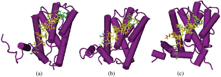
Structures of the Bcl-xL with three peptides for simulations at t=10 ns. (A: Bak-Bcl-xL system; B:HBS helix 2a-Bcl-xL system; C: HBS helix 2b-Bcl-xL system) Bcl-xL structures are shown in purple, residues in Bak from Gly572 to Gly575 and the corresponding cyclic rings in HBS helix 2a and HBS helix 2b are shown in green; residue Asp584 (KAC in HBS helix 2b) is shown in red, HBS helix 2b forms a Y shape and anchors at the hydrophobic surface of Bcl-xL. The rest Parts of the three inhibitive peptides are shown in yellow.