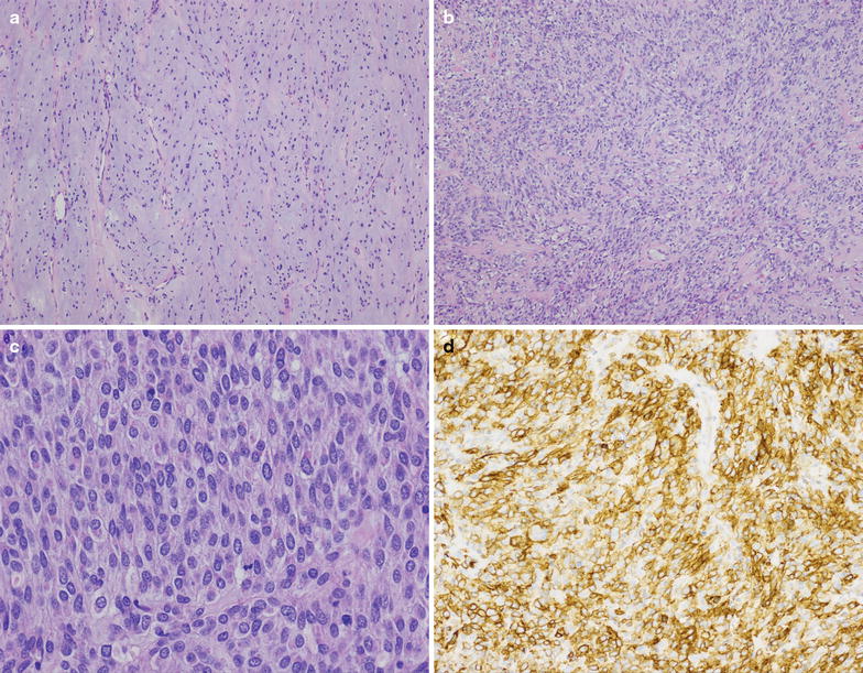
Fig. 1.

At low power (a, b, ×100), the metastatic GIST resected in 2014 demonstrated variable cellularity, with spindled to predominately epithelioid cells embedded within a myxoid stroma. On high power (c, ×400) cellular areas demonstrated sheets of epithelioid tumor cells with abundant cytoplasm and monotonous nuclei with finely granular chromatin, typical for epithelioid GIST. Scattered mitotic figures were present. Immunohistochemical study for DOG1 (d, ×200) was diffusely positive in tumor cells
