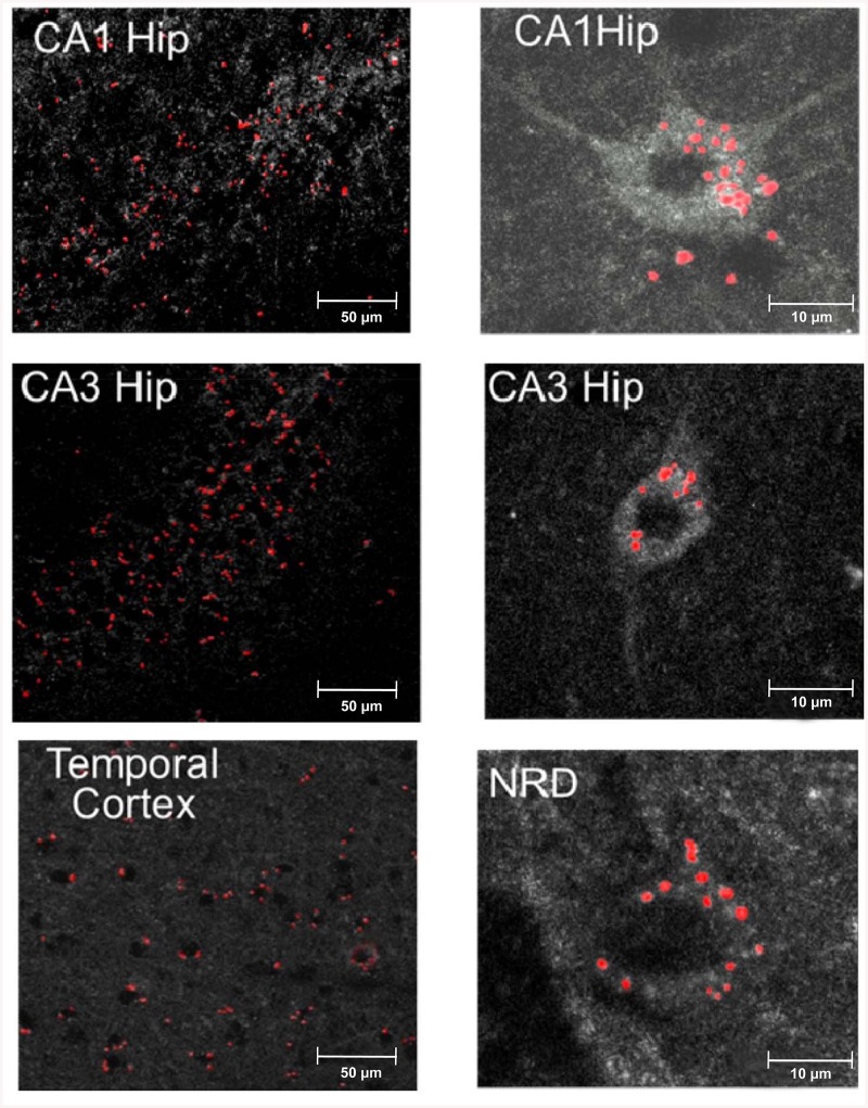
Fig 7. Penetration of intranasally-injected Cy3-labeled YB-1 into mouse brain regions.
Left, microphotographs of the temporal cortex and the hippocampal areas CA1 and CA3. Red spots represent labeled YB-1. Right microphotographs are examples of possible YB-1 penetration into neurons of CA1, CA3, and NRD (the serotonin-synthesizing nucleus raphe dorsalis).

