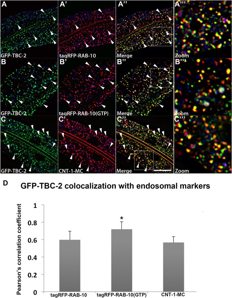
Fig 2. TBC-2 colocalizes with RAB-10 and CNT-1 on endosomes.
All images are from deconvolved 3D confocal image stacks acquired in intact living animals expressing GFP- and RFP-tagged proteins specifically in intestinal epithelial cells. (A-A'') GFP-TBC-2 colocalizes well with tagRFP-RAB-10. Arrowheads indicate endosomes labeled by both GFP-TBC-2 and tagRFP-RAB-10. (A‴) Magnified image of A'' is designated by rectangular outline. (B-B'') GFP-TBC-2 colocalizes extensively with tagRFP-RAB-10(Q68L), a predicted constitutively active form of RAB-10. Arrowheads indicate endosomes labeled by both GFP-TBC-2 and tagRFP-RAB-10(Q68L). (B‴) Magnified image of B'' is designated by rectangular outline. (C-C'') GFP-TBC-2 colocalizes well with CNT-1-MC. Arrowheads indicate endosomes labeled by both GFP-TBC-2 and CNT-1-MC. (C‴) Magnified image of C'' is designated by rectangular outline. In each image autofluorescent lysosome-like organelles appear in blue in all three channels, whereas GFP appears only in the green channel and RFP shows up only in the red channel. Signals observed in the green or red channels that do not overlap with signals in the blue channel are considered bona fide GFP or RFP signals respectively. (Scale bar: 10 μm) (D) Pearson's correlation coefficient for colocalization of GFP-TBC-2 with tagRFP-RAB-10, tagRFP-RAB-10(Q68L), and CNT-1-MC. n = 6. Error bars represent SEM. *P<0.05(student's t test).

