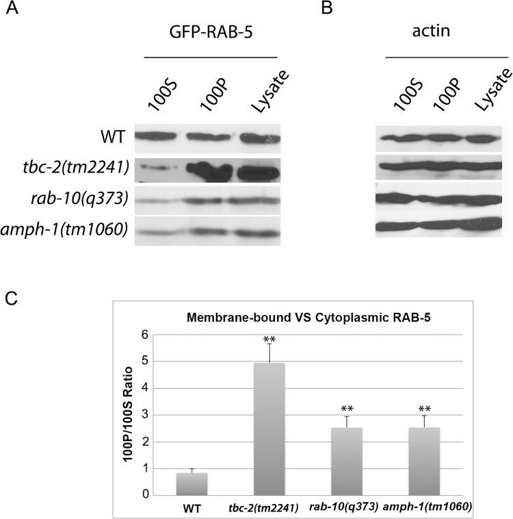
Fig 5. RAB-5 displays elevated membrane-association in tbc-2, rab-10 and amph-1 mutants.
(A) The membrane-to-cytosol ratio of RAB-5 increased in tbc-2, rab-10 and amph-1 mutants. Post-nuclear worm lysates were subject to centrifugation at 100,000g for 1 hour. 100P corresponds to the pellets and 100S represents the supernatants after the 100,000g centrifugation. (B) Loading control with actin antibody. (C) Quantification of the membrane-to-cytosol ratio (100P/100S) of RAB-5 in appropriate genetic backgrounds. The ratio of membrane-bound VS cytosolic RAB-5 was determined by densitometry. The standard deviations from three independent experiments are shown. **P<0.01 (student's t test).

