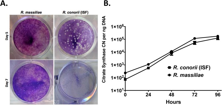
Fig 1. R. massiliae and R. conorii (ISF) infection resulted in plaques in Vero cell monolayers and replicated efficiently in the human dermal microvascular cell line, HMEC-1 cells.
(A) R. massiliae and R. conorii (ISF) were grown in Vero cells at 34°C for 5 and 7 days to quantify the number of plaques present as revealed by crystal violet staining (B). R. massiliae and R. conorii (ISF) were grown in HMEC-1 cells for times indicated. Data are representative of three independent experiments (A and B).

