
Fig 7. GFP-SseK3 localises to the trans-Golgi network.
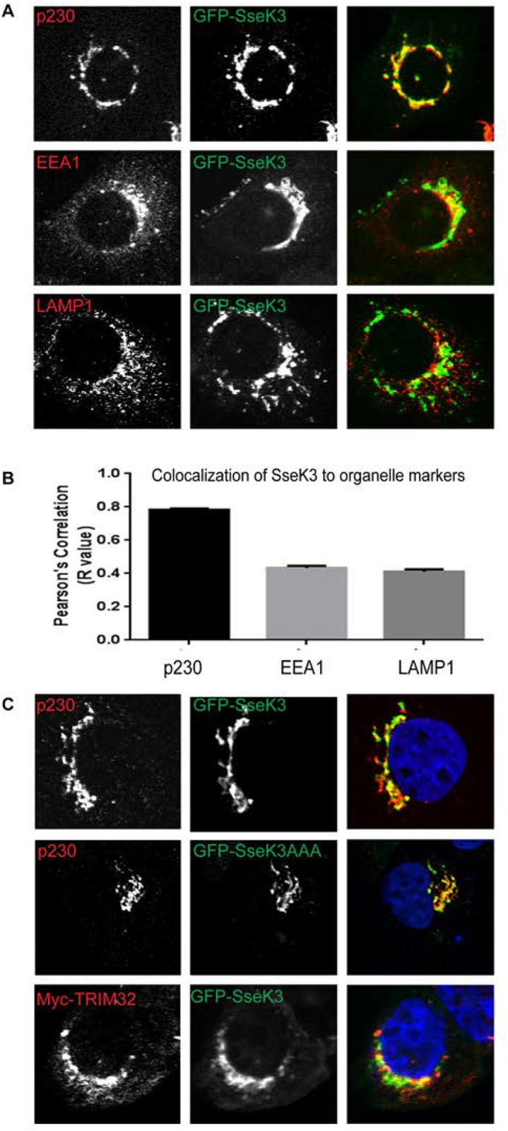
(A) Sub-confluent A431 cells grown on coverslips were transiently transfected with a plasmid encoding GFP-SseK3. 16–18 h post transfection, cells were fixed in 4% PFA and subjected to indirect immunofluorescence by staining with anti-p230, anti-EEA1 or anti-LAMP1 antibodies, followed by Alexa Fluor conjugated secondary antibodies. Images were captured using a Zeiss LSM 510 confocal laser-scanning microscope under 63x Oil objective. (B) Quantification of co-localisation between GFP-SseK3 and endogenous markers. Graph represents the mean Pearson’s Correlation (R value) of 100 cells randomly selected from 20 individual images. Error bars represent SEM. (C) A431 cells were transfected with plasmids encoding GFP-SseK3 or GFP-SseK3AAA alone, or Myc-TRIM32 and GFP-SseK3 together, and subjected to indirect immunofluorescence using anti-p230 and anti-Myc antibodies, followed by Alexa Fluor conjugated secondary antibodies. Images were captured using a Zeiss LSM 510 confocal laser-scanning microscope under 63x Oil objective. Scale bar 10μm.