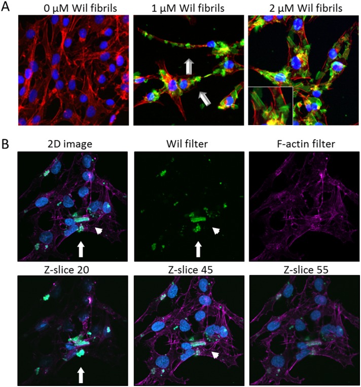
Fig 4. rVλ6Wil fibrils bind cultured cardiomyocytes.
(A) Fluorescent synthetic fibrils composed of rVλ6Wil (Alexa Fluor-488, green; arrow) bound specifically to cultured AC10 cardiomyocytes (blue, nuclei; red, f-actin). (B) Confocal micrographs of cultured cardiomyocytes (blue, nuclei; purple, f-actin) after incubation with 1 μM fluorescent rVλ6Wil fibrils (green). Optical sections at the surface (Z-slice 20), center (Z-slice 45), and bottom (Z-slice 55) demonstrated the presence of fibrils on the cell surface (arrow) and rare intracellular material (arrowhead). All images were enhanced equivalently by increasing the brightness.

