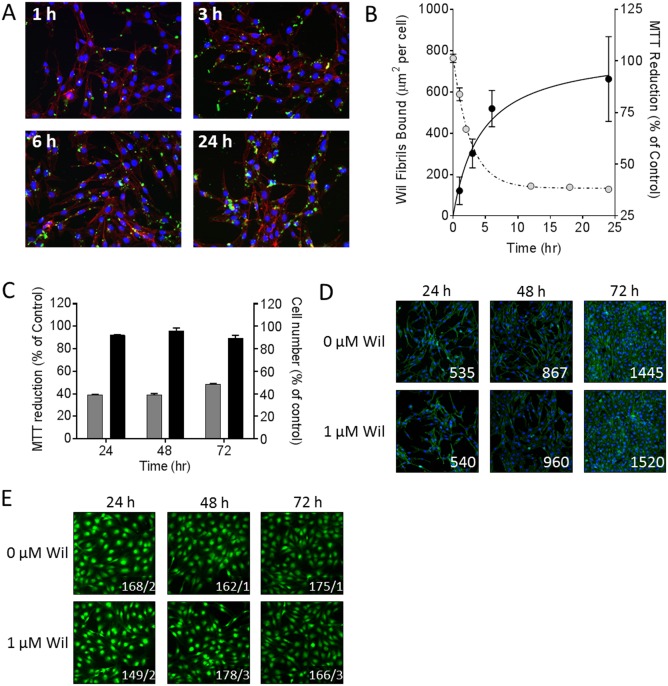
Fig 5. rVλ6Wil fibrils cause a decrease in cardiomyocytes MTT reduction without inducing cell death.
(A) Fluorescence microscopy demonstrating the increase in binding of Alexa Fluor-488 labeled rVλ6Wil fibrils (1 μM) over 24 h of incubation (blue, nuclei; red, f-actin). (B) Quantitation of rVλ6Wil fibril binding to cardiomyocytes (μm2/cell; closed circles) correlated inversely, over 24 h, with MTT reduction (open circles). (C) Cell number, quantified using crystal violet (black) did not decrease over 72 h of incubation with 1 μM rVλ6Wil fibrils despite a decrease in MMT reduction (grey; n = 3 samples per time point). (D) Fluorescence micrographs of AC10 cardiomyocytes incubated with or without non-fluorescent 1 μM rVλ6Wil fibrils (blue, nuclei; green, f-actin; numbers represent cell count in 10x objective field of view). (E) Cell viability of AC10 grown in culture for 4 days in the presence or absence of 1 μM rVλ6Wil fibrils for 24, 48, or 72 h, was assessed by CMFDA fluorescence (original objective, 20x. Images were cropped and digitally magnified 4x. Numbers represent the mean live/dead cells, n = 4 independent fields of view).

