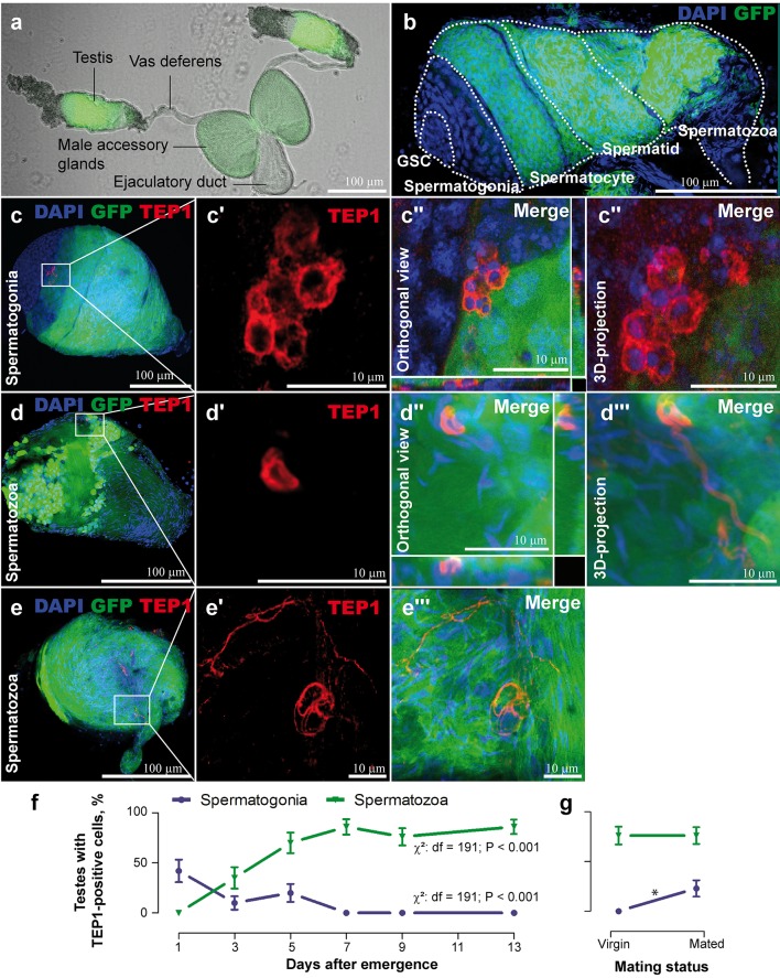
Fig 1. TEP1 occurrence in the testes during spermatogonial development.
The DSX transgenic line [12] that expresses GFP (green) under the β-tubulin promoter in the meiotic stages starting from spermatocytes but not in the mitotic germline stem cells (GSC) and spermatogonia. Nuclei were colored with DAPI (blue). (A) Male reproductive organs. (B) Organization of spermatogonial compartments in the testis (dotted lines). (C–E) TEP1 (red) recruitment to the spermatogonia (C–C”‘) and to the spermatozoa’s head (D–D”‘) and tail (E–E”). (F) Occurrence of testes with TEP1-positive cells during the first week after adult emergence. Testes were dissected for immunofluorescence analyses at the indicated time points. (G) Effect of mating on the percentage of testes with TEP1-positive cells. Virgin males (7-d-old) were collected in copula, and 2 d later their testes were dissected for immunofluorescence analyses. Significant differences (p < 0.05, χ2 test) are shown by an asterisk. Vertical bars show standard deviation, n ≥ 30 testes. Data used to make this figure can be found in S1 Data.

