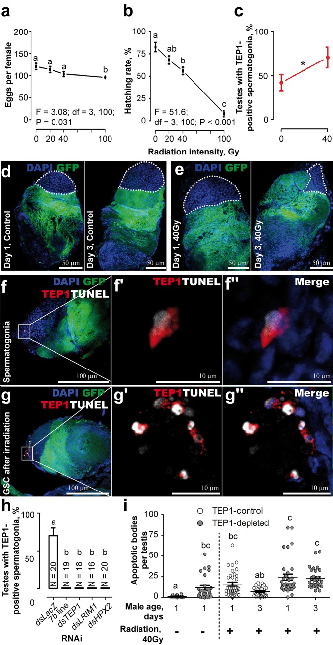
Fig 2. Effect of radiation on TEP1-mediated removal of damaged sperm.
A DSX transgenic line expressing GFP (green) under the β-tubulin promoter was used. Nuclei were colored with DAPI (blue). (A,B) 3-d-old virgin males that emerged from irradiated pupae were mated with 3-d-old virgin females, and the number of (A) laid eggs and the (B) larval hatching rates per female were gauged. Means ± standard error of the mean (SEM) are plotted for n ≥ 25. (C) The proportion of testes with TEP1-positive spermatogonia in irradiated males (40 Gy), n ≥ 30. (D,E) Radiation (40 Gy) reduces the size of the spermatogonial compartment (white dotted line) in 1- and 3-d-old males. (F,G) Colocalization of TEP1 (red) and TUNEL (white) signals in (F–F”) spermatogonia and (G–G”) the GSC in irradiated 1-d-old males. (H) Occurrence of TEP1 in spermatogonia of irradiated (40 Gy) males (DSX) injected with dsTEP1, dsLRIM1, dsHPX2, and dsLacZ (control). Males depleted for TEP1 (7b line) served as positive controls. The proportion of testes with TEP1 signal was gauged 2 d later. Mean ± standard error (SE) is shown; N, number of testes. (i) Accumulation of TUNEL-positive spermatogonia after irradiation in the testes of control and TEP1-depleted males (progeny of reciprocal crosses between 7b and DSX) was examined 1 and 3 d after emergence. Each dot represents one testis. Significant differences (p < 0.05, χ2 test) are shown by an asterisk and by characters above the corresponding values. Data used to make this figure can be found in S1 Data.

