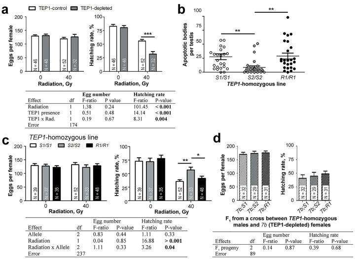
Fig 3. Effect of radiation and TEP1 depletion on male fertility.
Pupae were irradiated (40 Gy), and the resulting 3-d-old males were mated with 3-d-old females. The mean ± SEM of laid eggs and the proportions of hatched larvae are plotted. N, number of oviposited females. (A) After irradiation, TEP1 depletion (7b line) decreases hatching rates as compared to controls (T4 line). (B) The proportion of testes with apoptotic cells was examined by TUNEL staining in irradiated TEP1-homozygous (S1/S1, S2/S2, or R1/R1) 1-d-old males. Each dot represents one testis. (C) Irradiated TEP1-homozygous (S1/S1, S2/S2, or R1/R1) 3-d-old males were mated with TEP1*S1/S1 females. (D) TEP1 expression was silenced in the males of F1 reciprocal crosses between 7b and each of the TEP1-homozygogus lines. Irradiated F1 3-d-old males were mated with TEP1*S1/S1-homozygogus females. The results of two-way analysis of variance (ANOVA) tests are shown in tables below the corresponding graphs. Post hoc Tukey’s test: * p < 0.05; ** p < 0.01; *** p < 0.001. Data used to make this figure can be found in S1 Data.

