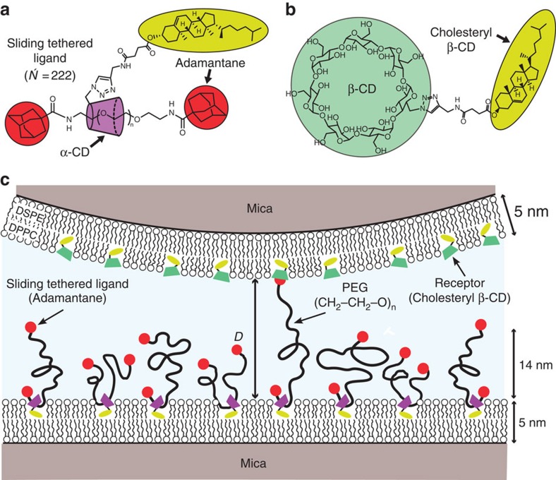
Figure 1. Experimental geometry.
(a) Molecular structure of a STL. (b) The cholesteryl β-CD receptor. (c) Molecular configuration used in the SFA experiments. Both membranes deposited on mica by a Langmuir–Blodgett technique were in a gel phase so as to minimize lateral mobility. The surface number density of STLs was σ=0.044 nm−2, which corresponds to 23 nm2 for each tethered ligand molecule (94:6 DPPC:STL). The STLs formed a polymer brush of mean thickness h0=14 nm, as determined from SFA compression curves and confirmed by neutron reflectivity experiments. Each STL is composed of a PEG polymer (N =222), which is threaded through the cavity of a cholesteryl α-CD membrane anchor and capped with adamantane at each end. There are on average 1.8 cholesteryl α-CD anchors per chain, as determined by nuclear magnetic resonance. In the opposing surface the ratio DPPC:cholesteryl β-CD is 90:10. The distance between opposing surfaces, D, is referred to and measured from the outer edge of the lipid head groups.

