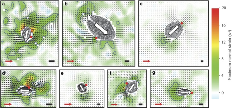
Figure 3.
Flow and shear fields of the seven species of dinoflagellates. Gyrodinium dominans (a); Akashiwo sanguinea (b); Dinophysis acuta (c); Oxyrrhis marina (d); Protoceratium reticulatum (e); Lingulodinium polyedrum (f); Amphidinium longum (g). Organisms have been cropped in from the original videos. White arrows indicate the swimming direction. Black length scales are 10 μm. Black vectors show the flow field, and the red arrow represents a 100 μm s−1 scale. The color scheme denotes the fluid deformation, and the blue contour line shows the 3 s−1 threshold value (see text). The red dots mark the point of prey capture, as either observed here or from the literature. See text for interpretation.

