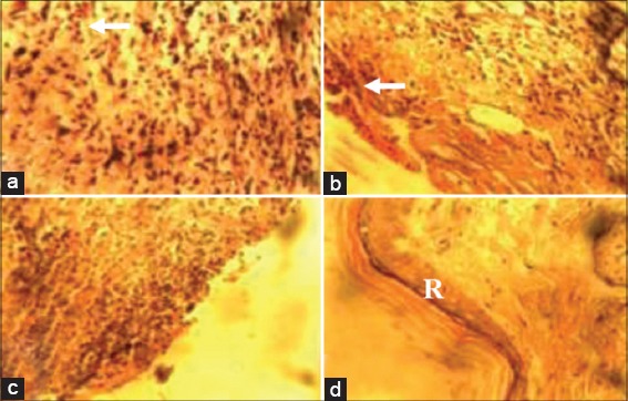
Figure 1.

Photomicrographs of wound site sections at day 7 post infliction of excision wound showing moderate inflammatory cell infiltrates (arrow) and more fibroblasts in wound sections of animals in Groups I (10% methanolic Crinum jagus bulb extract ointment [MCJBEO] treated) (a) and II (5% MCJBEO treated) (b), more inflammatory cell infiltrates in wound section of animals in Group IV (control) (c) and complete layer of epithelial regeneration (R) with more fibroblasts in wound section of animals in Group III (framycetin sulfate treated) (d). H and E ×400
