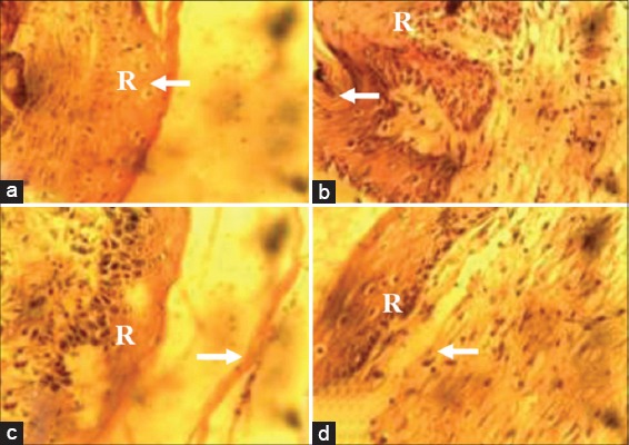
Figure 2.

Photomicrograph of wound site sections at day 14 post infliction of excision wound showing a complete layer of regenerated epithelium (R) with overlying keratin (arrow) which was greater in wound sections of animals in Groups I (10% methanolic Crinum jagus bulb extract ointment [MCJBEO] treated) (a), II (5% MCJBEO treated) (b) and III (framycetin sulfate treated) (c) than in wound sections of animals in the control group (d). H and E ×400
