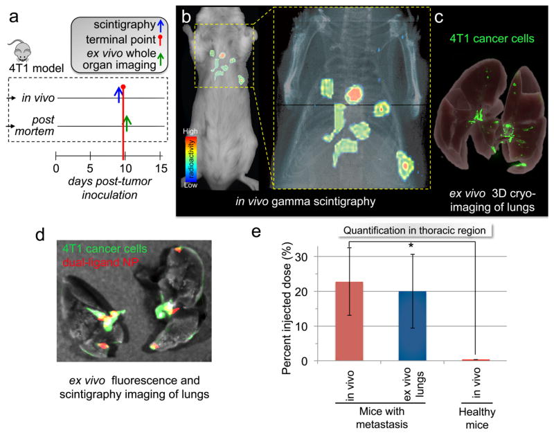
Figure 5. Targeting early-stage metastasis using the dual-ligand nanoparticle.
(a) The timeline and schedule is shown for the in vivo imaging studies and post mortem analyses. (b) Using whole-body planar gamma scintigraphy, representative coronal image shows the accurate targeting of a 99mTc-labeled dual-ligand-NP to early-stage metastases in the lungs of a mouse with 4T1 metastasis 2 h after injection. Following a systemic injection of ~20 μCi of 99mTc-dual-ligand-NP, the animals were imaged using a Gamma Medica X-SPECT system. The animal model was used 9 days after orthotopic tumor inoculation in a mammary fat pad, which was the time point of early onset of lung metastasis. (c) In the end of the in vivo imaging session, the lungs of the animals were perfused, excised and imaged ex vivo using planar gamma scintigraphy, planar fluorescence imaging and 3D cryo-imaging. 3D-cryo-imaging provided an ultra-high-resolution fluorescence volume of the lungs showing the early onset and topology of metastatic disease. (d) Overlaying ex vivo planar fluorescence and scintigraphy imaging of lungs, the colocalization of dual-ligand-NP and 4T1 metastatic cells is shown. (e) The targeting efficacy of dual-ligand-NP was compared in mice bearing 4T1 metastasis and healthy mice (n=4). Using a gamma scintigraphy, both groups of animals were imaged 2 h after systemic injection of ~20 μCi of 99mTc-dual-ligand-NP. The signal intensity in the thoracic region was compared quantitatively between the two groups.

