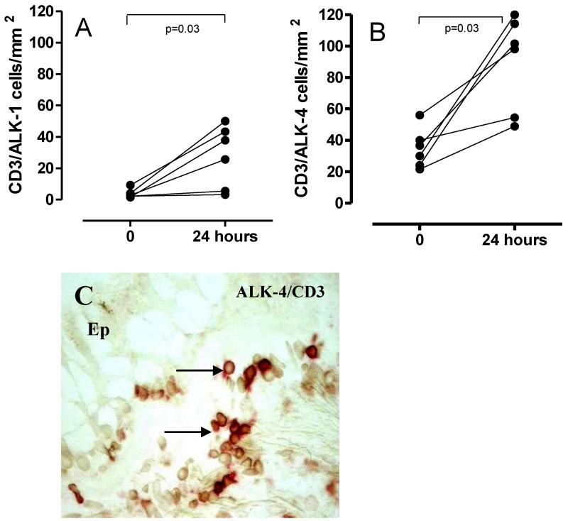
Figure 4. ALK-1 and ALK-4 modulation of expression CD3+ T cells post-allergen.
Numbers of T cells expressing receptors for TGFβ (ALK-1 Figure 4A) and activin (ALK-4, Figure 4B) were enumerated before and after allergen challenge by double staining for CD3 and ALK-1 or ALK-4. Counts are expressed as cells per mm2 of biopsy. A representative photomicrograph for double staining for CD3 (stained red) and ALK-4 (DAB) is shown in Figure 4C. Double stained cells are seen as a darker red-brown colour. The epithelium is indicated as Ep to orientate the section. N=6.

