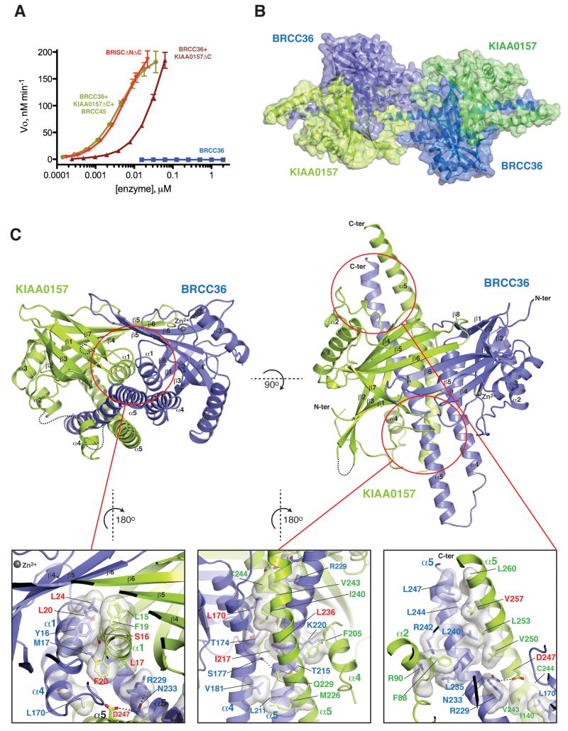
Figure 1. Structure of the BRCC36–KIAA0157 complex.
A) Cleavage activity of CfBRCC36 and CfBRCC36-containing complexes towards an internally quenched K63-diUb fluorogenic substrate. Results ± SEM are the average of three independent experiments carried out in duplicate.
B) Structure of the CfBRCC36–KIAA0157ΔC complex. Contents of the asymmetric unit revealed two CfBRCC36–KIAA0157ΔC complexes forming a dimer of heterodimers (super dimer).
C) Ribbon representation of the CfBRCC36–KIAA0157ΔC heterodimer. Zoom in panels show a detailed view of interacting residues. Disordered regions are shown as dashed lines. Interacting residues analyzed by mutation are labeled in red. See also Figures S1-S4 and S6.

