Figure 4.

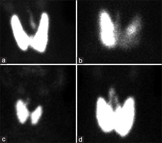
Variations in pyramidal lobe: (a) Arising from left lobe, (b) from isthmus, (c) from right lobe, (d) two pyramidal lobes joined by levator glandulae thyroideae (“inverted V”)

Variations in pyramidal lobe: (a) Arising from left lobe, (b) from isthmus, (c) from right lobe, (d) two pyramidal lobes joined by levator glandulae thyroideae (“inverted V”)