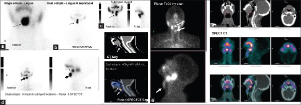
Figure 5.

99mTcO4 thyroid images showing different sites of ectopic thyroid tissue, (a) single ectopia at lingual location, (b) dual ectopia-at lingual and suprahyoid location with thyroid tissue along thyroglossal tract, (c) sublingual thyroid, (d) dual ectopia at hyoid and subhyoid location (planar and single photon emission computed tomography [SPECTCT] images) (e) planar TcO4 thyroid scan and SPECTCT images showing lingual thyroid (thin arrow) and thyroid tissue along thyroglossal tract (bold arrow) in the same patient, confirmed with SPECTCT images.
