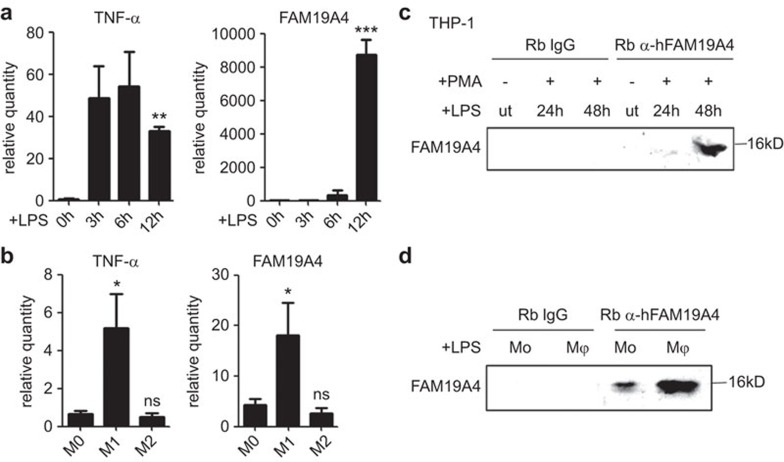
Figure 2.
The expression of FAM19A4 was inducible in the monocytes and macrophages. (a, c) The THP-1 cells were pre-treated with PMA (10 ng/ml) for 24 h and then stimulated with LPS (1 µg/ml). (a) FAM19A4 increased in the stimulated THP-1 cells at the mRNA level. The relative quantity is the ratio of 2−ΔΔCt compared to the control. (c) FAM19A4 increased in the cell culture medium of the stimulated THP-1 cells at the protein level. (b) FAM19A4 increased in the macrophages during their differentiation to M1 at the mRNA level. (d) FAM19A4 increased in the cell culture medium of the macrophages after LPS stimuli. The medium was first ultrafiltered and then immunoprecipitated using anti-FAM19A4 or normal rabbit IgG as the control (ut: untreated). The representative result of three independent experiments is shown.

