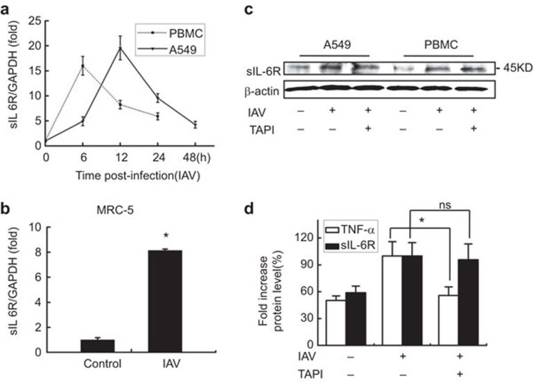
Figure 1.
IAV activates sIL-6R expression in different cell types. (a) sIL-6R mRNA levels were determined by real-time RT-PCR in A549 cells and human PBMCs infected with IAV (MOI=1) for the indicated length of time. (b) The level of sIL-6R mRNA was determined by real-time RT-PCR in MRC-5 cells infected with IAV (MOI=1) for 4 h. (c) sIL-6R protein levels were determined by western blot analysis of A549 cells and human PBMCs with (20 nM) or without TAPI for 8 h and were then infected with IAV (MOI=1) for 12 h. (d) A549 cells were incubated with (20 nM) or without TAPI for 8 h and were then infected with IAV (MOI=1) for 4 h. The levels of sIL-6R and TNF-α protein in the culture supernatants were measured by ELISA. Data shown are mean±s.e.; n=3. *P<0.05; ns, not significant. IAV, influenza A virus; MOI, multiplicity of infection; PBMC, peripheral blood mononuclear cell; sIL-6R, soluble interleukin-6 receptor; TNF, tumor-necrosis factor.

