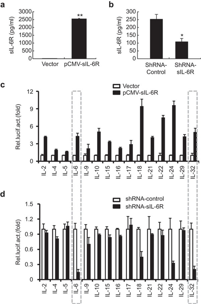
Figure 2.
Screen of IL-6R-stimulated cytokine promoter activity during IAV infection. (a) A549 cells were transfected with pCMV-Tag2B-sIL-6R for 24 h, and sIL-6R in the culture supernatants was quantitated by ELISA. (b) A549 cells were transfected with shRNA-sIL-6R or shRNA-control for 24 hand infected with IAV (MOI=1) for 6 h. The levels of sIL-6R protein in the culture supernatants were quantitated by ELISA. (c) Luciferase reporter plasmids for the indicated cytokines and a Renilla control (pRL–TK) were cotransfected into A549 cells with pCMV-Tag2B-sIL-6R or a control vector (pCMV–Tag2B) for 24 h. The luciferase activity was measured as described in the section on ‘Materials and methods'. (d) Luciferase reporter plasmids and a Renilla control (pRL–TK) were cotransfected with shRNA-sIL-6R or shRNA-control for 24 h into A549 cells that were infected with IAV (MOI=1) for 6 h, and the luciferase activity was measured. The results are expressed as the mean±s.e.m. of three independent experiments performed in triplicate and normalized according to the Renilla control reporter activity. n=3. *P<0.05. IAV, influenza A virus; sIL-6R, soluble interleukin-6 receptor; MOI, multiplicity of infection.

