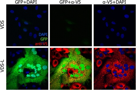
Figure 3.

Protein expression from PPRV-del-L after infection at higher moi. Cells were infected with PPRV-del-L at moi = 0.5. At 48 hours post infection, cells were fixed and stained as in Figure 2. Confocal images were taken by sequential scanning. Fields showing infection and protein expression in VDS and VDS-L are shown.
