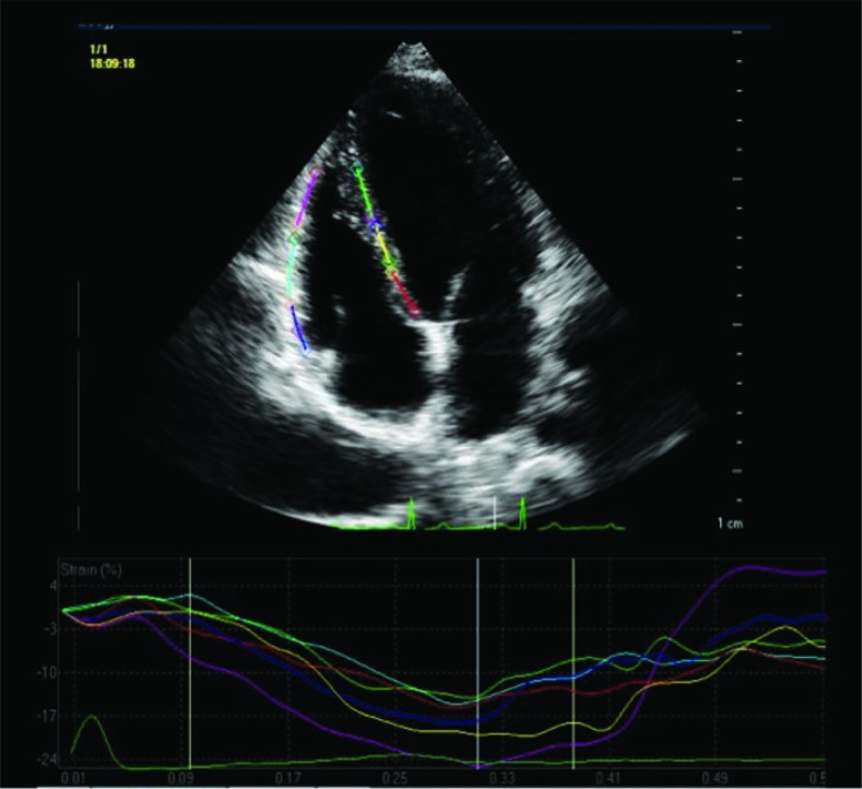
Fig. 3.
Echocardiography, apical 4-chamber view. Speckle tracking. Regions of interest. Below myocardial longitudinal strain curves determined by speckle tracking imaging
Legend: myocardial segments (regions of interest): dark blue line – basal segment of the lateral wall; red line – basal segment of the septum; blue line – middle segment of the lateral wall; yellow line – middle segment of the septum; purple line – apical segment of the lateral wall; green line – apical segment of the septum

